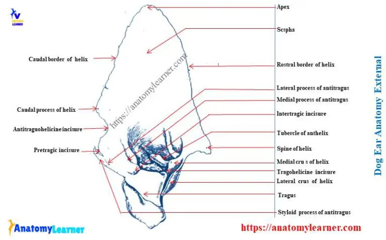
Dog Ear Anatomy Diagram

The canine ear is a complex and fascinating organ, responsible for detecting sound waves and maintaining balance. To understand the intricacies of dog ear anatomy, it’s essential to explore the various components that make up this remarkable structure.
External Ear Anatomy
The external ear, also known as the pinna or auricle, is the visible part of the dog’s ear. It’s composed of cartilage and skin, and its shape and size can vary greatly depending on the breed. The external ear is responsible for collecting sound waves and directing them into the ear canal.
- Pinna: The pinna is the outermost part of the ear, and it’s made of cartilage and skin. It’s responsible for collecting sound waves and funneling them into the ear canal.
- Ear Canal: The ear canal, also known as the external auditory meatus, is a narrow tube that connects the external ear to the eardrum. It’s lined with skin and cartilage, and it’s responsible for transmitting sound waves to the eardrum.
Middle Ear Anatomy
The middle ear is an air-filled cavity that contains the eardrum and the ossicles. The eardrum, also known as the tympanic membrane, is a thin, semi-transparent membrane that separates the ear canal from the middle ear.
- Eardrum (Tympanic Membrane): The eardrum is a thin, semi-transparent membrane that vibrates when sound waves reach it. These vibrations are then transmitted to the ossicles.
- Ossicles: The ossicles are three small bones (the malleus, incus, and stapes) that transmit vibrations from the eardrum to the inner ear. They’re responsible for amplifying sound waves and allowing the dog to hear a wide range of frequencies.
- Middle Ear Cavity: The middle ear cavity is an air-filled space that contains the eardrum and the ossicles. It’s connected to the back of the throat by the Eustachian tube, which equalizes air pressure on both sides of the eardrum.
Inner Ear Anatomy
The inner ear is a complex structure that’s responsible for detecting sound waves and maintaining balance. It’s composed of the cochlea, vestibule, and semicircular canals.
- Cochlea: The cochlea is a spiral-shaped structure that’s responsible for converting sound waves into electrical signals. It’s lined with thousands of hair cells that detect vibrations and transmit them to the brain.
- Vestibule: The vestibule is a small, fluid-filled sac that’s connected to the cochlea and the semicircular canals. It’s responsible for detecting changes in gravity and maintaining balance.
- Semicircular Canals: The semicircular canals are three small, fluid-filled tubes that are connected to the vestibule. They’re responsible for detecting rotational movements and maintaining balance.
Blood Supply and Innervation
The dog’s ear is supplied with blood by the external carotid artery and the internal carotid artery. The external carotid artery supplies the external ear, while the internal carotid artery supplies the middle and inner ear.
- External Carotid Artery: The external carotid artery supplies the external ear with oxygenated blood.
- Internal Carotid Artery: The internal carotid artery supplies the middle and inner ear with oxygenated blood.
- Facial Nerve: The facial nerve is responsible for controlling the muscles of the face, including the ear. It also transmits sensory information from the ear to the brain.
- Vestibulocochlear Nerve: The vestibulocochlear nerve is responsible for transmitting sensory information from the inner ear to the brain. It’s responsible for detecting sound waves and maintaining balance.
In conclusion, the dog ear anatomy is a complex and fascinating structure that’s responsible for detecting sound waves and maintaining balance. Understanding the various components of the dog’s ear can help us appreciate the intricacies of this remarkable organ and how it functions to allow dogs to hear and maintain their balance.
What is the function of the pinna in dog ear anatomy?
+The pinna, or external ear, is responsible for collecting sound waves and directing them into the ear canal. Its shape and size can vary greatly depending on the breed, and it's made of cartilage and skin.
What is the role of the ossicles in the middle ear?
+The ossicles are three small bones (the malleus, incus, and stapes) that transmit vibrations from the eardrum to the inner ear. They're responsible for amplifying sound waves and allowing the dog to hear a wide range of frequencies.
What is the function of the cochlea in the inner ear?
+The cochlea is a spiral-shaped structure that's responsible for converting sound waves into electrical signals. It's lined with thousands of hair cells that detect vibrations and transmit them to the brain.
As we’ve explored the intricacies of dog ear anatomy, it’s clear that this complex structure plays a vital role in a dog’s ability to hear and maintain balance. By understanding the various components of the dog’s ear, we can appreciate the remarkable complexity and beauty of this organ. Whether you’re a veterinarian, a dog owner, or simply someone fascinated by the intricacies of the canine ear, this journey into the world of dog ear anatomy has provided a unique and fascinating perspective on this remarkable structure.


