10+ Female Genitalia Diagrams For Accurate Knowledge

Understanding female genitalia is crucial for both medical professionals and the general public, as it promotes health, wellness, and sexual education. The female genital system is complex and multifaceted, consisting of both external and internal structures, each playing a significant role in reproductive and sexual health. To facilitate accurate knowledge, here are detailed descriptions and potential diagrams of the female genitalia, keeping in mind the importance of anatomical correctness and educational clarity.
External Female Genitalia
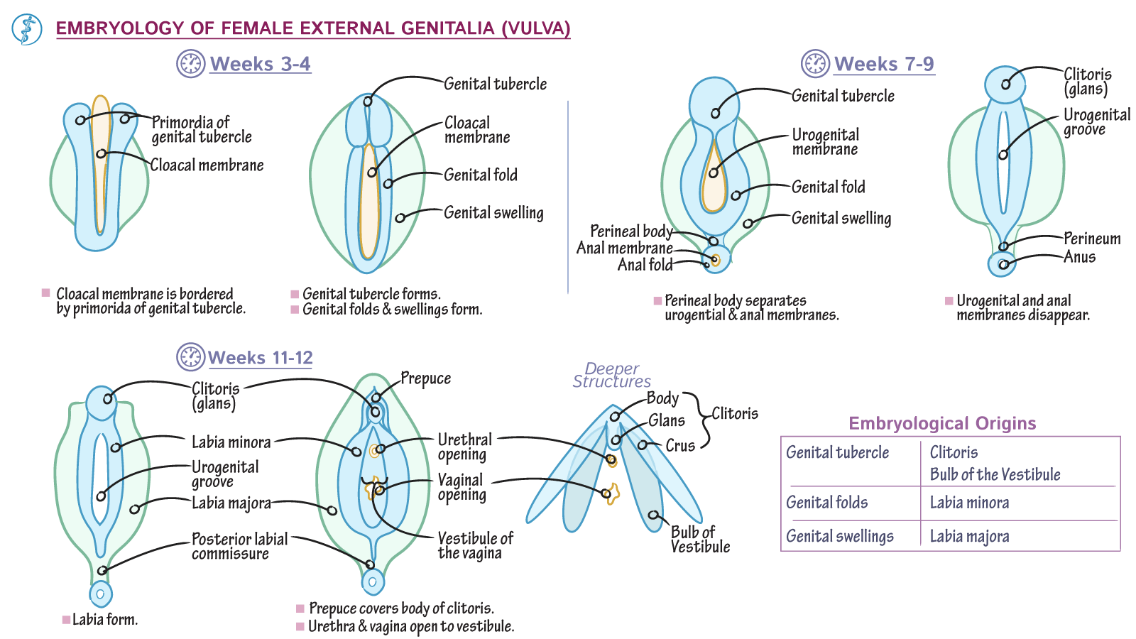
Vulva: The term “vulva” refers to the external parts of the female genitalia. It includes the labia majora, labia minora, clitoris, and the openings to the urethra and vagina.
- Labia Majora: These are the larger, fleshy folds of skin that protect the rest of the external genitalia. They contain sweat glands and sebaceous glands.
- Labia Minora: These are smaller, thinner folds of skin found within the labia majora. They surround the openings to the urethra and vagina and are highly sensitive.
- Clitoris: A highly sensitive organ that plays a significant role in female orgasm. It is located at the top of the vulva, where the labia minora meet.
- Urethral Opening: This is the opening through which urine leaves the body.
- Vaginal Opening: This is the opening of the vagina, which leads to the uterus.
Perineum: The perineum is the area between the vaginal opening and the anus. It is a crucial part of the pelvic floor and supports the pelvic organs.
Internal Female Genitalia
- Vagina: A muscular tube that connects the external genitals to the uterus. It serves as the passage through which menstrual fluid leaves the body and as the birth canal during delivery.
- Uterus (Womb): A hollow, muscular organ that supports fetal development during pregnancy. It is roughly the size of a fist and has three layers: the inner endometrium, the muscular myometrium, and the outer perimetrium.
- Ovaries: Two small, oval-shaped glands located on either side of the uterus. They produce eggs (oocytes) and hormones (estrogen and progesterone), which are crucial for reproductive health.
- Fallopian Tubes: These tubes connect the ovaries to the uterus, allowing eggs to travel from the ovaries to the uterus. Fertilization typically occurs within the fallopian tubes.
- Cervix: The lower part of the uterus, which opens into the vagina. It produces mucus, which changes in consistency throughout the menstrual cycle to either prevent or facilitate sperm entry.
Diagrams for Education
When referring to diagrams of female genitalia for educational purposes, it’s essential to ensure they are anatomically correct, detailed, and clear. Diagrams can include:
- Labeled Cross-Sections: Showing the layers of the vaginal wall or the structure of the uterus.
- 3D Models: Providing a comprehensive view of how the different structures are related spatially.
- Comparative Diagrams: Comparing the female genitalia at different stages of life, such as before and after puberty, or during pregnancy.
Importance of Accurate Representation
Accurate knowledge and representation of female genitalia are crucial for several reasons: - Health Awareness: Understanding the anatomy helps in identifying abnormalities or issues early on. - Sexual Education: Correct knowledge promotes healthy sexual relationships and practices. - Empowerment: Knowledge about one’s own body is a form of empowerment, allowing individuals to make informed decisions about their health and well-being.
In conclusion, diagrams and detailed descriptions of female genitalia are essential tools for education and awareness. They provide a visual aid that can help in understanding the complex structures and their functions, fostering a deeper appreciation and respect for the human body.
